产品详情
组分(Materials Provided)
IDComponentsSizeRSP001-C01Pre-coated Anti-IFN-γ Antibody Microplate1 plateRSP001-C02Positive Stimulus10 μgRSP001-C03Biotin-Anti-IFN-γ Antibody50 μLRSP001-C04Streptavidin-HRP50 μLRSP001-C05Washing Buffer (10x)50 mLRSP001-C06Dilution Buffer (1x)50 mLRSP001-C07AEC Dilution25 mLRSP001-C08AEC Solution A (20x)0.8 mLRSP001-C09AEC Solution B (20x)0.8 mLRSP001-C10AEC Solution C (20x)0.8 mL产品概述(Product Overview)
Interferon-γ (IFN-γ) is the only type II interferon. This proinflammatory cytokine is secreted by activated T cells and NK cells. It activates macrophages and endothelial cells and regulates immune responses by affecting APCs, T cells, and B cells. Production of IFN-γ by helper T cells and cytotoxic T cells is a hallmark of the Th1-type phenotype. Thus, high-level production of IFN-γ is typically associated with effective host defense against intracellular pathogens. The Human IFN-γ ELISpot assay is designed for the detection of human IFN-γ secreting cells at the single cell level and can be used to quantitate the frequency of human IFN-γ secreting cells. ELISpot assays are highly reproducible and sensitive, and can be used to measure responses with frequencies well below 1 in 100,000. ELISpot assays do not require prior in vitro expansion of T cells and they are suitable for high-throughput analysis using only small volumes of primary cells. As such, ELISpot assays are useful tools for research in vaccine development, cytokine secretion and the monitoring of various clinical trials.
应用说明(Application)
This kit is used to detect IFN-γ at the cellular level.
It is for research use only.
重构方法(Reconstitution)
Please see Certificate of Analysis for details of reconstitution instruction and specific concentration.
存储(Storage)
The unopened kit is stable for 12 months from the date of manufacture if stored at 2°C to 8°C.
原理(Assay Principles)
ELISpot assays employ the quantitative sandwich enzyme-linked immunosorbent assay (ELISA) technique. The Kit consists of Pre-coated Anti-IFN-γ Antibody Microplate, Positive Stimulus, Biotin-Anti-IFN-γ Antibody, Streptavidin-HRP, AEC and buffers.
Your experiment will include 6 simple steps:
a) A monoclonal antibody specific for human IFN-γ has been pre-coated onto a polyvinylidene difluoride (PVDF)-backed microplate.
b) Appropriately stimulated cells are pipetted into the wells and the microplate is placed into a humidified 37 °C CO2 incubator for a specified period of time.During this incubation period, the immobilized antibody in the immediate vicinity of the secreting cells binds secreted IFN-γ.
c) After washing away any cells and unbound substances, a biotinylated antibody specific for human IFN-γ is added to the wells. Following a wash to remove any unbound biotinylated antibody, HRP conjugated to streptavidin is added.
d) Wash the plate and add the Streptavidin-HRP diluted by Dilution Buffer to the plate.
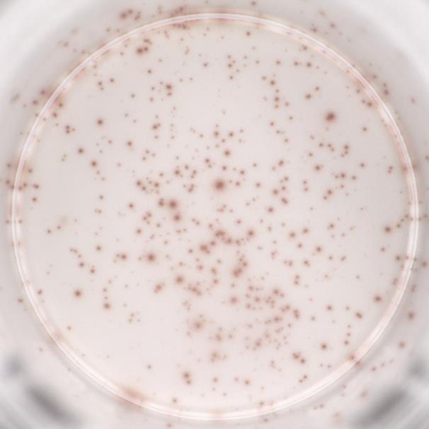
e) Wash the plate and add AEC. A reddish-brown colored precipitate forms and appears as spots at the sites of cytokine localization, with each individual spot representing an individual IFN-γ secreting cell.
f) SThe spots can be counted with an automated ELISpot reader system or manually using a stereomicroscope.
质量管理控制体系(QMS)
产品展示
典型数据-Typical Data
Please refer to DS document for the assay protocol.

5x10⁴ cells/well PBMC were stimulated with 0.2 μg/mL Positive Stimulus and cultured at 37℃ and 5% CO2 for 20 h, 328 spots were produced. The following example data is for reference only.
用户评价 发表评论

