产品详情
抗体来源(Source)
Monoclonal Anti-G4S linker Antibody, Rabbit IgG (016) is a rabbit monoclonal antibody recombinantly expressed from human 293 cells (HEK293), which provides higher batch consistency and long term security of supply. It shows superior performance in flow cytometry assays with higher affinity and better specificity.
应用(Application)
Flow Cytometry (Evaluation of cell surface expressed CARs of varying specificity containing a G4S linker within the scFv of the extracellular domain).
克隆号(Clone)
016
种属(Species)
Rabbit
亚型(Isotype)
Rabbit IgG | Rabbit Kappa
特异性(Specificity)
Specifically recognizes the scFv-based CARs containing a G4S linker.
偶联(Conjugate)
PE
Excitation Wavelength: 488 nm / 561 nm
Emission Wavelength: 575 nm
推荐稀释比(Recommended Dilution)
1:50
制剂(Formulation)
Lyophilized from 0.22 μm filtered solution in PBS, 0.03% Proclin 300, pH7.4, 0.2% BSA with trehalose as protectant.
Contact us for customized product form or formulation.
重构方法(Reconstitution)
Please see Certificate of Analysis for specific instructions.
For best performance, we strongly recommend you to follow the reconstitution protocol provided in the CoA.
存储(Storage)
Please protect from light and avoid repeated freeze-thaw cycles.
This product is stable after storage at:
- -20°C to -70°C for 24 months in lyophilized state;
- -70°C for 12 months after reconstitution.
- 2-8°C for 12 months after reconstitution.
质量管理控制体系(QMS)
产品展示
活性(Bioactivity)-FACS
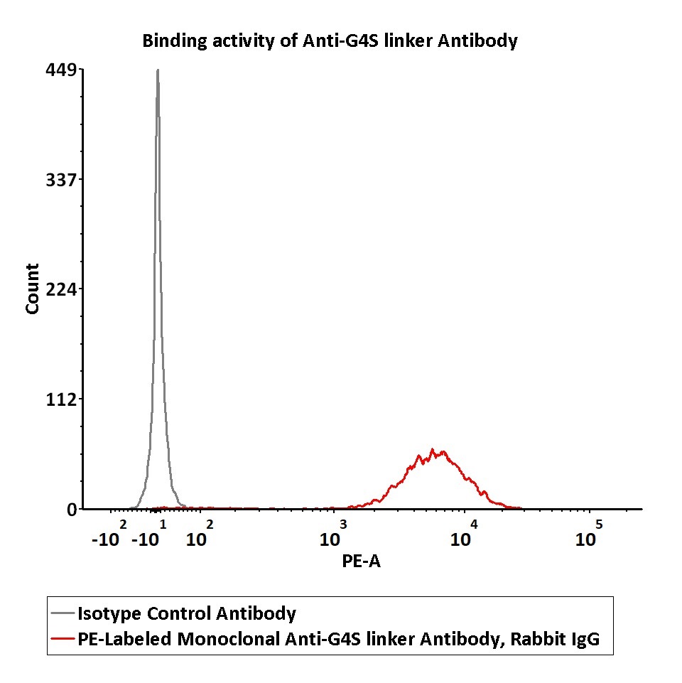
Flow cytometric analysis of Anti-MSLN CAR-293 cells staining with PE-Labeled Monoclonal Anti-G4S linker Antibody, Rabbit IgG (016) (Cat. No. G4S-PFMY25) at 1:50 dilution (2 μL of the antibody stock solution corresponds to labeling of 1e6 cells in a final volume of 100 µL), compared with isotype control antibody. PE signal was used to evaluate the binding activity (QC tested).
Protocol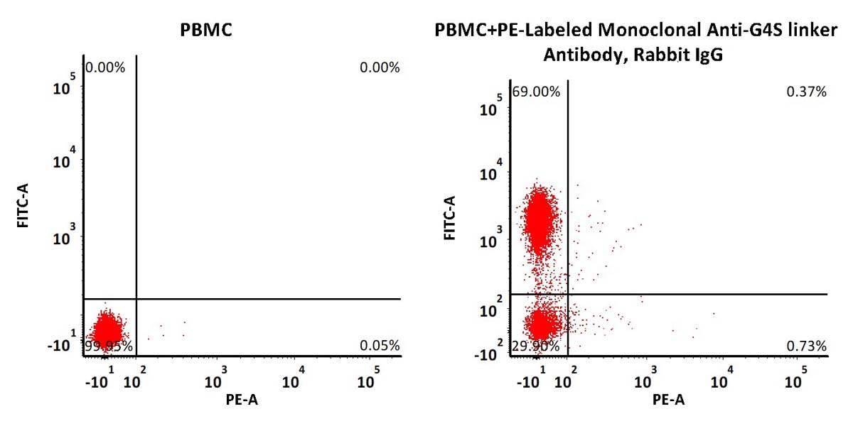
Non-specificity of PE-Labeled Monoclonal Anti-G4S linker Antibody, Rabbit IgG (016) (Cat. No. G4S-PFMY25) binding to CD3+ cells present in human PBMC. 5e5 of human PBMCs were simultaneously stained with FITC-labeled anti-CD3 antibody and PE-Labeled Monoclonal Anti-G4S linker Antibody (2 μL of the antibody stock solution corresponds to labeling of 5e5 cells in a final volume of 100 µL) and washed and then analyzed with FACS. Both FITC and PE positive signals was used to evaluate the non-specific binding activity to human CD3+ cells (QC tested).
Protocol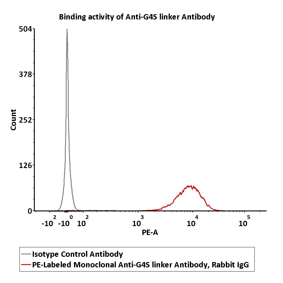
Flow cytometric analysis of Anti-CD19 CAR-293 cells staining with PE-Labeled Monoclonal Anti-G4S linker Antibody, Rabbit IgG (016) (Cat. No. G4S-PFMY25) at 1:50 dilution (2 μL of the antibody stock solution corresponds to labeling of 1e6 cells in a final volume of 100 µL), compared with isotype control antibody. PE signal was used to evaluate the binding activity (Routinely tested).
Protocol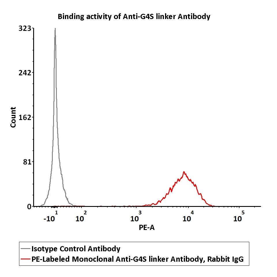
Flow cytometric analysis of Anti-CD22 CAR-293 cells staining with PE-Labeled Monoclonal Anti-G4S linker Antibody, Rabbit IgG (016) (Cat. No. G4S-PFMY25) at 1:50 dilution (2 μL of the antibody stock solution corresponds to labeling of 1e6 cells in a final volume of 100 µL), compared with isotype control antibody. PE signal was used to evaluate the binding activity (Routinely tested).
Protocol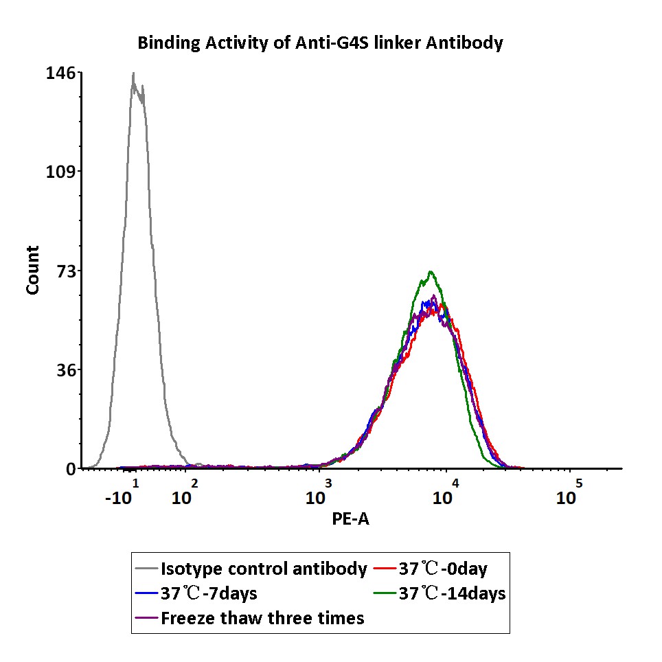
PE-Labeled Monoclonal Anti-G4S linker Antibody, Rabbit IgG (016) (Cat. No. G4S-PFMY25) is stable at 37℃ for 14 days or after freezing and thawing 3 times.
Protocol
Compared Data
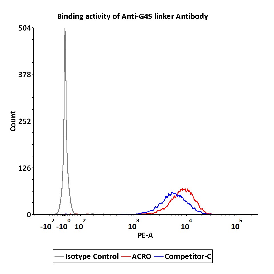
Flow cytometric analysis of Anti-CD19 CAR-293 cells staining with PE-Labeled Monoclonal Anti-G4S linker Antibodies. PE signal was used to evaluate the binding activity of anti-G4S linker antibody. The biological activity level of ACRO is superior to competitor C (Routinely tested).
Protocol
Non-specificity of PE-Labeled Monoclonal Anti-G4S linker Antibody, Rabbit IgG1 (016) (Cat. No. G4S-PFMY25) binding to CD3+ cells present in human PBMC. ACRO shows only 0.37% non-specificity signal, much lower than competitor C (9.78%).
Protocol
用户评价 发表评论

